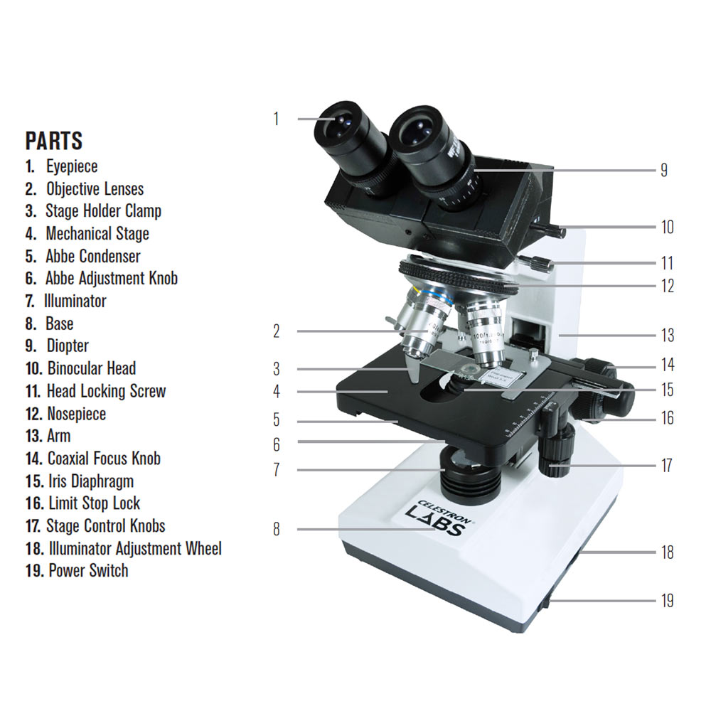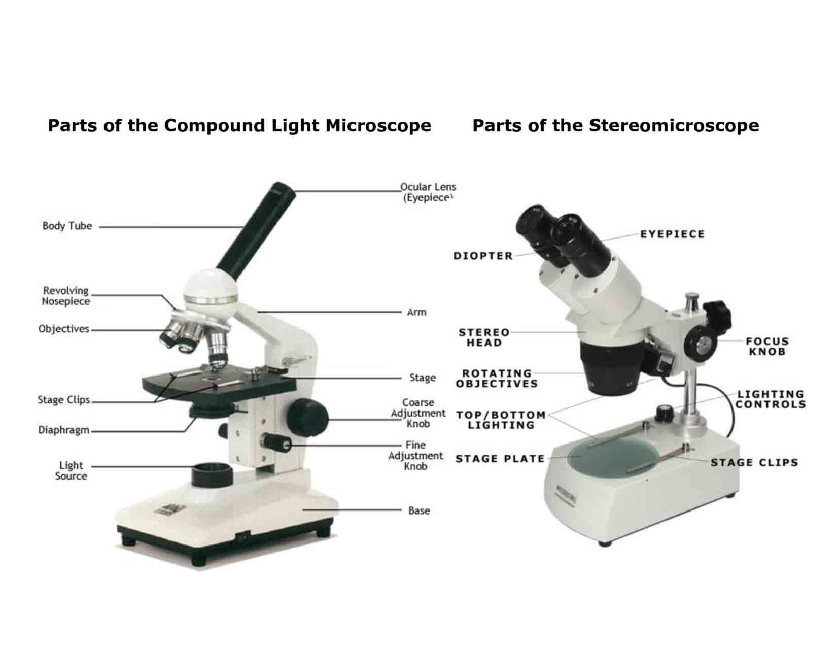
Together, the optical and mechanical components of the microscope, including the mounted specimen on a glass micro slide and coverslip, form an optical train with a central axis that traverses the microscope base and stand. The optical components contained within modern microscopes are mounted on a stable, ergonomically designed base that allows rapid exchange, precision centering, and careful alignment between those assemblies that are optically interdependent. Light leaving the objective is diverted by a beamsplitter/prism combination either into the eyepieces to form a virtual image, or straight through to the projection lens mounted in the trinocular extension tube, where it can then form a latent image on film housed in the camera system. The condenser forms a cone of illumination that bathes the specimen, located on the microscope stage, and subsequently enters the objective. Also stationed in the microscope base is a series of filters that condition the light emitted by the incandescent lamp before it is reflected by a mirror and passed through the field diaphragm and into the substage condenser. Illumination is provided by a tungsten-halogen lamp positioned in the lamphouse, which emits light that first passes through a collector lens and then into an optical pathway in the microscope base. Presented in Figure 1 is a typical microscope equipped with a trinocular head and 35-millimeter camera system for recording photomicrographs.

The intensity of illumination and orientation of light pathways throughout the microscope can be controlled with strategically placed diaphragms, mirrors, prisms, beamsplitters, and other optical elements to achieve the desired degree of brightness and contrast in the specimen. Most microscopes provide a translation mechanism attached to the stage that allows the microscopist to accurately position, orient, and focus the specimen to optimize visualization and recording of images. Modern compound microscopes are designed to provide a magnified two-dimensional image that can be focused axially in successive focal planes, thus enabling a thorough examination of specimen fine structural detail in both two and three dimensions.

Microscope Optical Components Introduction Molecular Expressions Microscopy Primer: Anatomy of the Microscope - Microscope Optical Components


 0 kommentar(er)
